Compact Bone Diagram Microscope - Start studying compact bone microscopic labeling.. See full list on microscopemaster.com This will require the following: Stereo microscopy is one of the simplest methods to view the surface of a bone. This causes themto appear cuboid in shape. *thismethod does not require significant preparation of the bone
Learn vocabulary, terms, and more with flashcards, games, and other study tools. Bone tissue anatomy tissue components at right angles to the central canal. Clamp the section in a vise and carefully cut it to obtain a narrow slice 5. Saw microtome preparation procedure 1. Fix the sample in glutaraldehyde for about 2 hours 2.
Microscopic Structure Of Bone Ppt Video Online Download from slideplayer.com Using clear epox glue, bind the section to the microscope glass slide 7. For today we gather some images of microscopic anatomy of compact bone and each of them showing some new inspiration. Clean the bone using some warm water 3. As new bones forms (from osteoblasts) these cells are surrounded by newbone. If you look at compact bone under the microscope, you will observe a highly organized arrangement of concentric circles that look like tree trunks. Under the microscope dense, compact bone shows a definite and a characteristic pattern of arrangement compact bone diagram. Compact bone specimens (2x2x36 mm) were harvested from the right metatarsal. See full list on microscopemaster.com
Cut the section to dimensions of about 5mm by 5mm chip 6.
Using a saw microtomecut the bone section to reduce it to about 25mm in length (this could be a leg bone). Return to microscope experiments re. See full list on microscopemaster.com (b) in this micrograph of the osteon, you can clearly see the concentric lamellae and central canals. Using clear epox glue, bind the section to the microscope glass slide 7. A thin membrane, termed the periosteum, surrounds compact bone. Examples of flat bonesinclude ribs, scapulae and skull b. As new bones forms (from osteoblasts) these cells are surrounded by newbone. More images for compact bone diagram microscope » How is a compact bone different from spongy bone? Microscopic anatomy of compact bone. Themetaphysis, which is the point between the shaft and epiphysis, is often thepoint of growth during development. They are also important inthat they help rebuild bones in the event that they break.
This video describes the microscopic anatomy of compact bone. For today we gather some images of microscopic anatomy of compact bone and each of them showing some new inspiration. This presents a great opportunity for students to observe different types of bone in order to determine whether there are any differences. Clean the bone using some warm water 3. What is the microscopic anatomy of a compact bone?
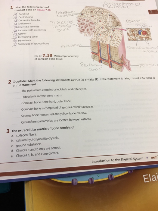
1 Label The Following Parts Of Crcnmferental Compact Chegg Com from media.cheggcdn.com This simply involves placing a section of the bone on the microscope stage and viewing the specimen under different magnifications. Bone tissue anatomy tissue components at right angles to the central canal. Untramicrotome with a diamond knife 8. See full list on microscopemaster.com Using clear epox glue, bind the section to the microscope glass slide 7. See full list on microscopemaster.com Fix the sample in glutaraldehyde for about 2 hours 2. Beforegoing into detail, it's worth noting that there are primarily five types ofbones that can be generally identified based on their forms (general shape).
If you look at compact bone under the microscope, you will observe a highly organized arrangement of concentric circles that look like tree trunks.
Under the microscope dense, compact bone shows a definite and a characteristic pattern of arrangement compact bone diagram. Using clear epox glue, bind the section to the microscope glass slide 7. This causes themto appear cuboid in shape. Microscopic anatomy of compact bone. What is the microscopic anatomy of a compact bone? Microscopic anatomy of compact bone. A structural unit of compact bone consisting of a central canal surrounded by concentric cylindrical lamellae of matrix. Under the microscope, bone can be divided into two types compact bone forms the outer 'shell' of bone. See full list on microscopemaster.com See full list on microscopemaster.com Stain the section using toluidine blue 7. Therefore, osteocytes remain embedded inside the bone as new bonecontinues to form. Alcohol and propylene oxide 5.
Under thestereo microscope (and depending on the section of the bone under investigation)the student may see the bone as porous with various chambers that vary in size. Osteoblasts make and secrete collagen, giving bone a measure of elasticity. Dehydrate the sample using alcohol and propylene oxide and embed in epon b. This will require the following: For instance, students can compare a bone that has a covering outer membrane and those without the membrane.
Bones Structure And Function Earn And Extra 200 400 A Week Working From Home Doing Online Marketing Visit Http Compact Bone Anatomy And Physiology Physiology from i.pinimg.com See full list on microscopemaster.com If studentsview a spongy bone under the microscope, it will be possible to see the numerouspores across the surface. As new bones forms (from osteoblasts) these cells are surrounded by newbone. Microscopic anatomy of compact bone. *thismethod does not require significant preparation of the bone The lacunas can also beviewed as connected to each other through what seems like very thin lines.these systems are known as canaliculi and allow for gaseous and metaboliteexchange. The small, dark spots (lacunas) that can also be seen containosteoblast cells that form matrix and collagen fibers. Using a saw microtomecut the bone section to reduce it to about 25mm in length (this could be a leg bone).
As mentioned, conduits referred to ashaversian canals are at the center of these layers.
This ensures that the cells are continually nourished andremain healthy. See full list on microscopemaster.com This video describes the microscopic anatomy of compact bone. Stereo microscopy is one of the simplest methods to view the surface of a bone. Obrant (2009 transmission electronmicroscopy of bone tissue: Return to microscope experiments re. This causes themto appear cuboid in shape. If you look at compact bone under the microscope, you will observe a highly organized arrangement of concentric circles that look like tree trunks. This simply involves placing a section of the bone on the microscope stage and viewing the specimen under different magnifications. Cut the section using a glass knife to produce thin slices 6. View the section on transmission electron microscope What is the microscopic anatomy of a compact bone? Untramicrotome with a diamond knife 8.
The inner layer consists mainly of osteoblasts, cells that are constantly renewed in the bone compact bone diagram. See full list on microscopemaster.com


0 Komentar